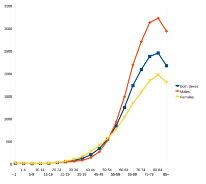
Atrophic gastritis
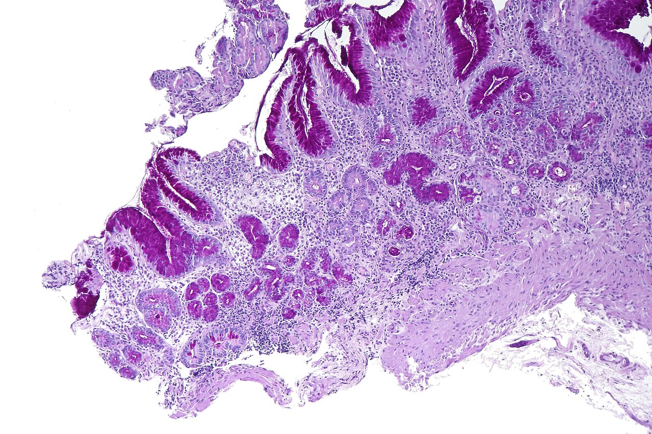
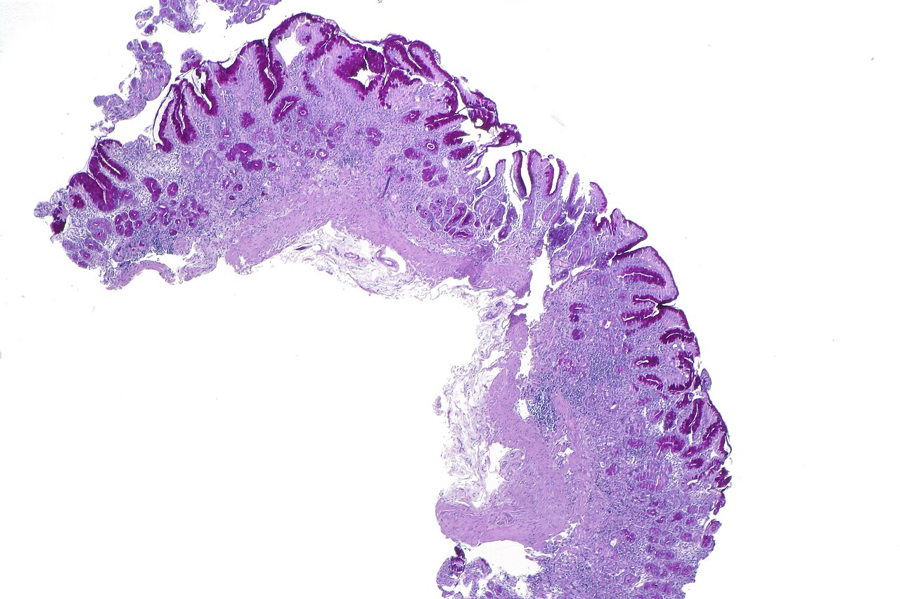
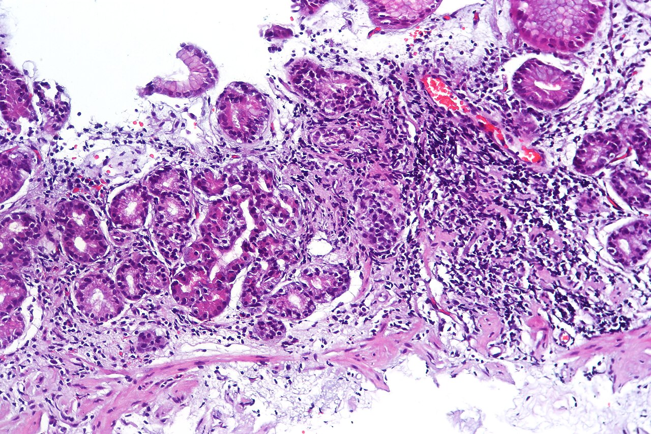
Chronic stomach inflammation causing loss of glandular cells.

CoRus13 · CC BY-SA 4.0
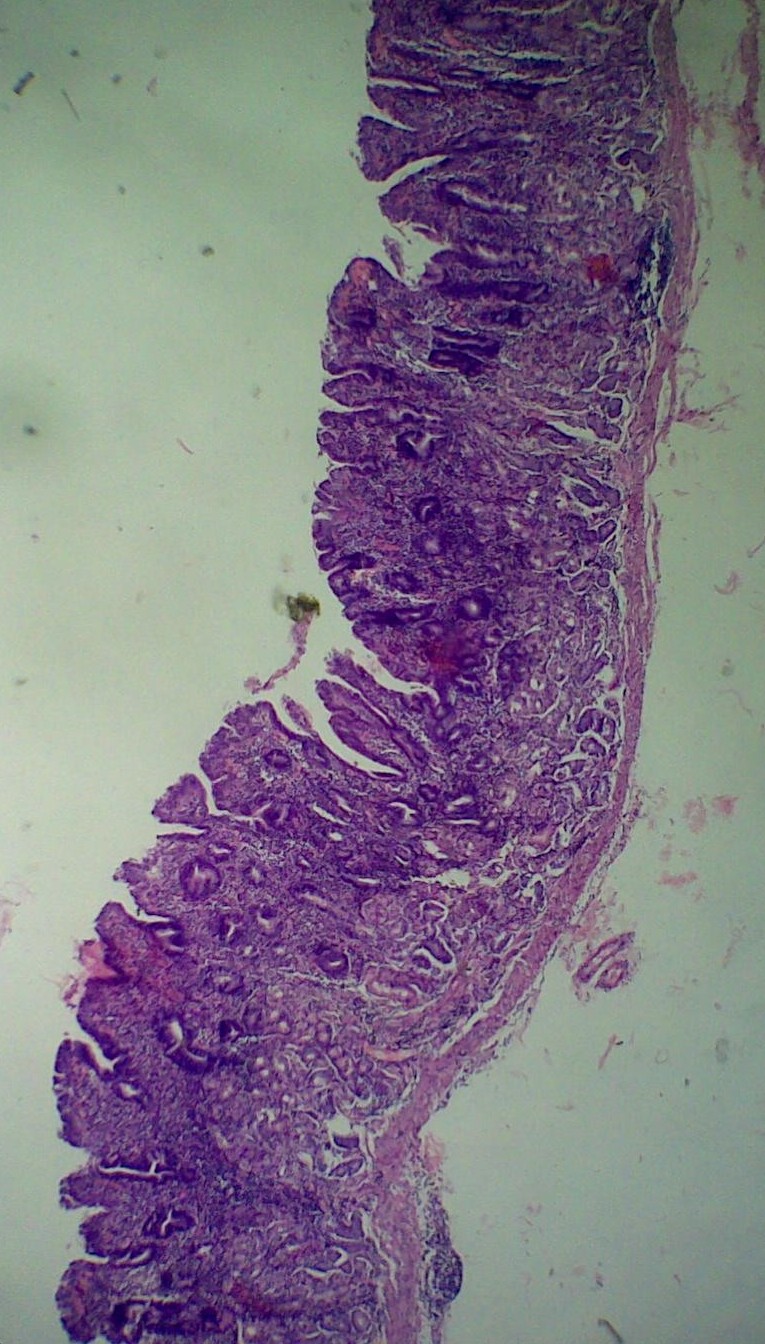
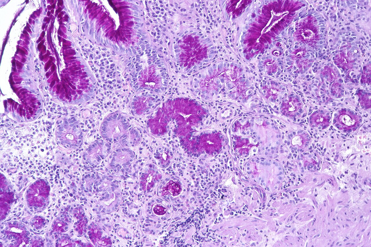
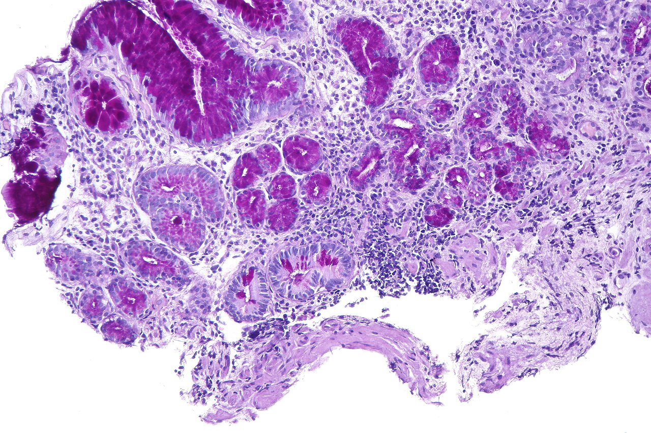
Atrophic gastritis is a chronic inflammatory condition of the stomach lining that leads to the loss of gastric glandular cells and their replacement by intestinal and fibrous tissues. This impairs the secretion of essential substances such as hydrochloric acid, pepsin, and intrinsic factor, resulting in digestive problems including pernicious anemia and iron deficiency anemia.
- diagnosis
- Detection of APCA, IFA, and Helicobacter pylori antibodies with serum gastrin
- treatment
- Supplementation of vitamin B12 and iron in deficient patients
Lore & Background
Atrophic gastritis is classified into two main types: Type A (autoimmune metaplastic atrophic gastritis) and Type B (environmental metaplastic atrophic gastritis). Type A primarily affects the fundus of the stomach and is more common with pernicious anemia, while Type B primarily affects the antrum and is more common with H. pylori infection. Autoimmune atrophic gastritis results from the immune system attacking parietal cells, leading to achlorhydria and hypergastrinemia, which may contribute to malignant transformation of enterochromaffin-like cells into carcinoid tumors.
Reader's Guide
Atrophic gastritis is significant as a precursor to serious conditions such as gastric adenocarcinoma and pernicious anemia. The condition impairs absorption of iron and vitamin B12, with iron deficiency often detected at a younger age than pernicious anemia. Patients with autoimmune atrophic gastritis are statistically more likely to develop gastric carcinoma, Hashimoto's thyroiditis, and achlorhydria. Diagnosis relies on detection of specific antibodies and serum gastrin levels. Supplementation of folic acid in deficient patients has been shown to improve histopathological findings and reduce the incidence of gastric cancer. The condition may be asymptomatic in some, but symptomatic patients are mostly female, presenting with iron deficiency signs and less commonly with neurological or psychiatric disorders.
Did You Know?
- Atrophic gastritis can be caused by persistent Helicobacter pylori infection or by autoimmune processes.
- Autoimmune atrophic gastritis impairs iron and vitamin B12 absorption, with iron deficiency detected at a younger age than pernicious anemia.
- Patients with autoimmune atrophic gastritis are statistically more likely to develop gastric carcinoma, Hashimoto's thyroiditis, and achlorhydria.
- Supplementation of vitamin B12 and iron in deficient patients can improve symptoms and reduce the risk of complications.
Pathophysiology & the Cascade of Cellular Loss
Atrophic gastritis begins as a persistent inflammatory process within the stomach's mucosal lining, gradually eroding the specialized glandular cells responsible for producing hydrochloric acid, pepsin, and intrinsic factor. Over time, these lost cells are replaced by intestinal-type and fibrous tissue, fundamentally altering the organ's digestive capacity. In the autoimmune variant, known as autoimmune metaplastic atrophic gastritis, the body's own immune system directs a sustained attack against parietal cells and intrinsic factor, a process that appears to carry an inherited component. The resulting absence of acid production, termed achlorhydria, triggers a compensatory cascade: gastrin-producing G cells undergo hyperplasia, elevating circulating gastrin levels. This excess gastrin exerts a trophic, growth-stimulating influence on enterochromaffin-like cells, which normally secrete histamine. Researchers hypothesize that this chronic stimulation may drive the malignant transformation of these cells into carcinoid tumors, representing one pathway by which autoimmune atrophic gastritis can progress toward neoplasia.
Clinical Presentation & the Spectrum of Symptoms
Many individuals living with atrophic gastritis experience no overt symptoms whatsoever, making the condition difficult to detect without targeted screening. When symptoms do manifest, they disproportionately affect women and often mirror the hallmarks of iron deficiency: persistent fatigue, restless legs syndrome, brittle fingernails, noticeable hair loss, a weakened immune response, and slower wound healing. A substantial proportion of symptomatic patients, roughly eighty percent, report delayed gastric emptying, while about a quarter experience reflux complaints or peripheral neuropathy. Rarer manifestations, occurring in one to two percent of cases, include autonomic nervous system abnormalities and progressive memory loss. The psychiatric landscape is equally concerning, with documented associations to mania, depression, obsessive-compulsive disorder, psychosis, and broader cognitive impairment. Notably, although both iron and vitamin B12 absorption are compromised in the autoimmune form, iron deficiency tends to surface at a younger age than the pernicious anemia that characterizes advanced B12 depletion, offering an earlier clinical window for detection.
Etiology, Subtypes & Classification Debates
Atrophic gastritis is broadly divided into two principal forms based on etiology and anatomical distribution. Type A, or autoimmune metaplastic atrophic gastritis, targets the fundus and body of the stomach and is strongly linked to pernicious anemia. It arises when the immune system mounts a sustained assault on parietal cells, a process with clear hereditary undertones. Type B, or environmental metaplastic atrophic gastritis, predominantly involves the antrum and is most frequently driven by persistent Helicobacter pylori infection or dietary factors; it is typically confined to the stomach's body region. Both variants carry an elevated risk of progressing to gastric adenocarcinoma, though the autoimmune form additionally predisposes patients to Hashimoto's thyroiditis and complete achlorhydria. Despite early proposals to categorize the disease by progression stage into so-called closed or open types, no single classification system has achieved universal acceptance in the medical literature as of 2017, reflecting ongoing debate about how best to stratify this heterogeneous condition.
Diagnostic Markers & Therapeutic Strategies
Diagnosing atrophic gastritis relies on a combination of serological and laboratory markers rather than a single definitive test. Clinicians look for anti-parietal cell antibodies, anti-intrinsic factor antibodies, and evidence of Helicobacter pylori exposure through antibody testing, all interpreted alongside serum gastrin levels to paint a coherent diagnostic picture. The absence of a universally accepted staging classification means that treatment decisions often depend on the individual patient's deficiency profile. For patients identified as deficient in folic acid, supplementation has been shown to improve the histopathological appearance of the chronically inflamed mucosa and, importantly, to reduce the long-term incidence of gastric cancer. Beyond targeted supplementation, management centers on correcting the downstream consequences of impaired gastric secretion, most critically the vitamin B12 deficiency that drives pernicious anemia and the iron malabsorption that produces a separate anemia. Ongoing surveillance for gastric adenocarcinoma remains a cornerstone of care, particularly for those with the autoimmune subtype.
Gallery
Frequently Asked Questions
Who is Atrophic gastritis?
Atrophic gastritis is a chronic inflammatory condition of the stomach lining that progressively destroys gastric glandular cells and replaces them with intestinal-type and fibrous tissue. It functions as a slow, persistent adversary to the stomach's structural integrity.
What are Atrophic gastritis's powers or role?
Its core ability is shutting down the stomach's production of hydrochloric acid, pepsin, and intrinsic factor. This cascade of lost secretions ultimately triggers pernicious anemia and iron deficiency anemia in affected patients.
How does Atrophic gastritis's story end?
The glandular cell loss itself is largely irreversible, so the narrative shifts to long-term management. Patients with documented deficiencies are treated with vitamin B12 and iron supplementation to stabilize their condition and prevent further systemic harm.
Why is Atrophic gastritis important in the broader canon?
It acts as a critical early-warning marker, signaling that the stomach's protective and digestive machinery is failing. Catching it before advanced damage occurs allows clinicians to intervene with targeted supplementation and monitoring.
How do you identify Atrophic gastritis in the field?
Diagnosis hinges on detecting autoantibodies (APCA and IFA), Helicobacter pylori antibodies, and elevated serum gastrin levels in a patient's bloodwork. Together, these serological markers confirm the condition's presence and likely etiology.
More in Aging-associated diseases 1-24
Spotted an error? Know more?
This is a living reference — every entry is fact-audited, and reader corrections feed straight into our audit queue. Suggest an edit · See this site's audit record